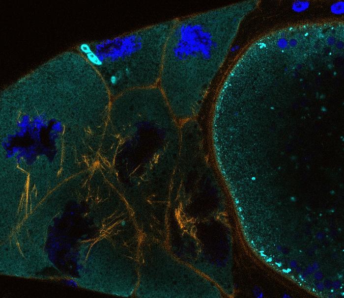
Image caption: Drosophila nurse cells and oocyte, actin (orange), cytoplasm (cyan), nuclei (blue).
Coordinated change and programmed cell death in small cell networks
Supervisor: Professor Tim Weil
Small cell networks coordinate collective change by reading and responding to cues. These decisions must be highly regulated in both time and space, especially during animal development. Examples of spatiotemporal regulation include cell contraction initiating tissue morphogenesis, clustering of migratory cells, and programmed cell death.
The overall aim of this project is to understand the mechanisms that underpin coordinated cell changes. As our model system, we use the experimentally attractive Drosophila nurse cells, which produce the mRNA and proteins essential for oogenesis and early embryogenesis. These cells collectively transfer their cytoplasm into the developing egg and then undergo synchronised death. Previous work has shown that these processes require a conserved signal from a group of surrounding epithelial cells as well as kinase activity and an intact actomyosin network.
Building on preliminary data in the lab suggesting that nurse cell volume change is asynchronous and a temporal intracellular calcium rise, we will interrogate the mechanism coordinating the extrusion of cytoplasm and subsequent death. We will use live cell imaging, advanced genetic manipulation, and modelling to test different working models in the field. Initial experiments may include targeted mutagenesis of candidate genes with CRISPR/Cas9, the visualisation of genetically encoded calcium sensors, and mosaic analysis of mutant nurse cells. Outcomes of this project will broadly inform our understanding of support cells in animal development and the regulation of cell networks which is implicated in pathogenesis and the design of cancer therapeutics.
For more information please visit WeilLabCambridge.com or contact Tim Weil via tw419@cam.ac.uk.
References
Imran Alsous J, et al., Dynamics of hydraulic and contractile wave-mediated fluid transport during Drosophila oogenesis. Proc Natl Acad Sci U S A. 2021;118(10).doi: 10.1073/pnas.2019749118.
Weil TT, Parton RM, and Davis I., Preparing individual Drosophila egg chambers for live imaging. J Vis Exp. 2012; (60).